
Illustration.
The Science of Muse Cells
What Muse cells are, where they live in the body, how scientists identify them, and the four behaviors that make researchers interested in them.
What Muse Cells Are
Muse cells, short for multilineage-differentiating stress-enduring cells, are naturally occurring stem cells. Scientists describe them as pluripotent-like: in lab and animal studies they can form many different cell types, yet unlike embryonic stem cells they have not formed tumors called Teratoma A usually benign tumor containing a jumble of tissue types, such as hair, muscle and bone. Fully pluripotent cells can form one if they are implanted before being turned into a specific cell type. in animal testing.1,39,41 Whether truly pluripotent cells exist in adult tissue is still debated in the wider field.45
Muse cells have been found in bone marrow, blood, skin, fat and the connective tissue of many organs.1,4,5,9,11,39 Umbilical cord tissue is another promising source being studied.7,8
They are rare. In bone marrow, by the most common counting method, roughly 1 in every 2,500 to 10,000 mononuclear cells is a Muse cell (about 0.01 to 0.04 percent).1,12,39 They make up roughly 1 percent to a few percent of standard Mesenchymal stem/stromal cells (MSCs) Stromal cells grown from tissues such as bone marrow, fat or umbilical cord. They mainly form bone, cartilage and fat, and release healing signals. cultures.3,39
After a stroke or heart attack, the number of Muse cells in the blood goes up. In one study, heart attack patients with bigger rises recovered heart function better. This suggests Muse cells may be part of the body’s own repair system, but cause and effect are not yet proven.12,13
How Scientists Find Them
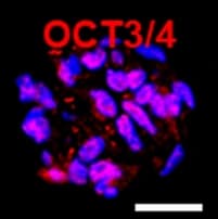
Scientists identify Muse cells mainly by a surface marker called SSEA-3 Stage-specific embryonic antigen 3, a sugar-based marker on the cell surface. Scientists use it to find and sort Muse cells., usually together with CD105, a marker they share with mesenchymal stem cells.1,2,3,11,43
There are three common ways to collect them from a culture:
- Sorting cells one by one by their fluorescent markers (FACS), the most common method, and it gives high purity.
- Pulling them out with magnetic beads that stick to SSEA-3 (MACS).
- Leaving the culture in trypsin, a digestive enzyme, for a long time, which enriches the stress-tolerant cells.
Methods and yields still vary between laboratories, and researchers are working toward standard protocols.1,3,7,28,43
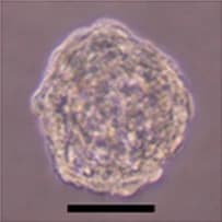
Evidence: Laboratory (stage 1 of 4)What Muse Cells Actually Look Like
Real microscope images from published, openly licensed studies, shown at their true size. Unlike the illustrations elsewhere on this site, these are data.
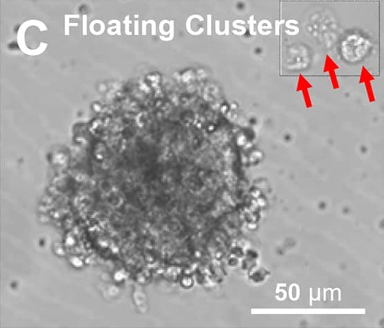
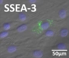
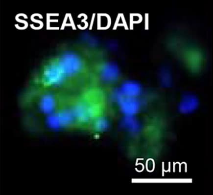
Four Things Researchers Have Observed
Each behavior, what the studies found, and how far the evidence has reached.
They Endure Stress
In lab studies, Muse cells survived harsh conditions and DNA-damaging stress better than neighboring cells, and repaired DNA damage efficiently.15 They also release protective proteins, including Serpins Serine protease inhibitors, a family of proteins that switch off protein-cutting enzymes. They help control inflammation, clotting and cell survival. and 14-3-3 proteins A family of small helper proteins inside cells that regulate many processes, including whether a stressed cell survives or self-destructs..14 Researchers think this toughness may help them survive in injured tissue, but that has not been measured in people.
Evidence: Laboratory (stage 1 of 4)They Follow a Damage Signal
Injured tissue releases a signaling molecule called S1P Sphingosine-1-phosphate, a small fat-based signaling molecule released by damaged cells. It can act like a distress flare.. In rabbit and mouse studies, Muse cells sensed S1P through a receptor called S1PR2 Sphingosine-1-phosphate receptor 2, a sensor on the cell surface that detects S1P. In animal studies, it guides Muse cells toward injury. and gathered at the injury, and blocking that receptor reduced this homing.16,17 In mouse models of stroke and ALS, Muse cells injected into a vein also reached the injured brain or spinal cord.22,23
In heart attack patients, blood Muse cell counts rose along with blood S1P levels, which is a correlation, not proof of cause.13 Human trials have not yet tracked where injected cells go.39
Evidence: Animal studies (stage 2 of 4)They Become What the Tissue Needs
In lab studies, single Muse cells can form cell types from all three Three germ layers The three basic tissue layers of the early embryo (ectoderm, mesoderm and endoderm). They give rise to every organ, from skin and nerves to muscle, bone and gut., a result reproduced by independent labs.1,5,6,10 In animal studies, Muse cells that reached an injury took on the features of the local cell type, for example liver or heart muscle cells.16,24,25
One proposed explanation is that Muse cells take up fragments of dying cells and use their signals to become the same kind of cell. Scientists are still working out how this happens.20
Evidence: Laboratory and animal studies (stage 2 of 4)They May Avoid Rejection
Many Muse cells carry HLA-G An immune-signaling molecule, best known from the placenta, that helps calm the immune system so the mother does not attack the fetus., a molecule the placenta uses to help protect the fetus from the mother’s immune system. How much HLA-G they carry varies with the tissue they come from.8,40
In the CL2020 trials so far, donor Muse cells were given without tissue matching or anti-rejection drugs, and no serious rejection reactions were reported.34,39 How long donor cells survive in the body, and why they seem to avoid rejection, are still open questions: in mice with normal immune systems, human Muse cells were rejected within 7 weeks.26 Mesenchymal stem cells were long called immune privileged too, and are now described as immune evasive.51
Evidence: Early human trials (safety observations only) (stage 3 of 4)
What About Tumors?
In animal studies, Muse cells have not formed tumors, and they show low activity of telomerase, an enzyme linked to unlimited cell growth. In the small human trials published so far, no tumor has been attributed to Muse cells. Longer follow-up in larger groups is still needed.1,4,8,22,38,39
How Muse Cells Compare
Muse cells, mesenchymal stem cells (MSCs) and induced pluripotent stem (iPS) cells each have real strengths. Muse cells are a distinct, promising research area, not a replacement for the others.
| Question | Muse cells | MSCs | iPS cells |
|---|---|---|---|
| What they are39,49,47 | A small subgroup (about 1 percent to several percent) of MSC and fibroblast cultures, identified by the marker SSEA-3 | A mixed population of stromal cells grown from bone marrow, fat, umbilical cord and other tissues | Adult cells reprogrammed in the lab to an embryonic-like state using defined genes |
| Found naturally in the body?1,11,49,47 | Yes: in bone marrow, blood and connective tissue | Obtained by growing cells from tissue in the lab | No: created in the lab |
| Genetic reprogramming needed?1,47 | No | No | Yes (reprogramming factors) |
| Range of cell types they can form1,5,10,45,49,39,47 | “Pluripotent-like”: cell types from all three germ layers in lab and animal studies, reproduced by several labs. Whether adult tissues hold truly pluripotent cells is still debated in the field. | Mainly bone, cartilage and fat; a broader range is debated | Fully pluripotent: cell types from all three germ layers |
| Tumor risk1,39,47,48 | No teratomas in animal studies and low telomerase activity; no tumor attributed to Muse cells in the small CL2020 trials so far | Generally considered low for adult somatic stem cells | Undifferentiated iPS cells form teratomas, so they are turned into the needed cell type before use; a Kyoto trial saw no graft overgrowth in 7 patients |
| After intravenous injection16,23,50,48 | In animals, they travel to injured tissue; not yet tracked in people | In animals, many are trapped in the lungs on their first pass through the body | Usually implanted directly at the target (for example, into the brain in Parkinson’s trials) |
| Immune handling of donor cells34,39,51,74 | CL2020 trials used unmatched donor cells without anti-rejection drugs; how long donor cells survive in people is unknown | Described as “immune evasive, not immune privileged” | Donor grafts in the Kyoto Parkinson’s trial were given with an anti-rejection drug (tacrolimus) |
| Clinical stage53,52,48,32,33,34,35,36,38,39 | Early trials in Japan (57 people treated with CL2020 in published studies); no approved product | Approved products exist, for example FDA-approved remestemcel-L (Ryoncil) for children with steroid-refractory acute graft-versus-host disease (December 2024) | Early-phase trials, for example a phase 1/2 trial in Parkinson’s disease |
Glossary
- SSEA-3
- Stage-specific embryonic antigen 3, a sugar-based marker on the cell surface. Scientists use it to find and sort Muse cells.
- HLA-G
- An immune-signaling molecule, best known from the placenta, that helps calm the immune system so the mother does not attack the fetus.
- S1P
- Sphingosine-1-phosphate, a small fat-based signaling molecule released by damaged cells. It can act like a distress flare.
- S1PR2
- Sphingosine-1-phosphate receptor 2, a sensor on the cell surface that detects S1P. In animal studies, it guides Muse cells toward injury.
- Serpins
- Serine protease inhibitors, a family of proteins that switch off protein-cutting enzymes. They help control inflammation, clotting and cell survival.
- 14-3-3 proteins
- A family of small helper proteins inside cells that regulate many processes, including whether a stressed cell survives or self-destructs.
- Pluripotency
- The ability of a cell to become any of the body’s cell types, from all three germ layers. “Pluripotent-like” means showing much of this ability in lab and animal tests.
- Three germ layers
- The three basic tissue layers of the early embryo (ectoderm, mesoderm and endoderm). They give rise to every organ, from skin and nerves to muscle, bone and gut.
- Allogeneic
- From a different person of the same species: donor cells rather than the patient’s own (which are called autologous).
- Teratoma
- A usually benign tumor containing a jumble of tissue types, such as hair, muscle and bone. Fully pluripotent cells can form one if they are implanted before being turned into a specific cell type.
- Mesenchymal stem/stromal cells (MSCs)
- Stromal cells grown from tissues such as bone marrow, fat or umbilical cord. They mainly form bone, cartilage and fat, and release healing signals.
- iPS cells
- Induced pluripotent stem cells: adult cells reprogrammed in the lab into an embryonic-like state.
- Homing
- The movement of cells through the bloodstream to a particular site, such as an injury.
- CL2020
- The development code for the donor Muse cell product made by Life Science Institute, Inc. and used in the Japanese clinical trials. All published human trial results for Muse cells come from this one product.
Last reviewed against its sources: . How we check claims
Have a Question About Muse Cell Research?
Dr. Albert’s team will read it and reply by email.